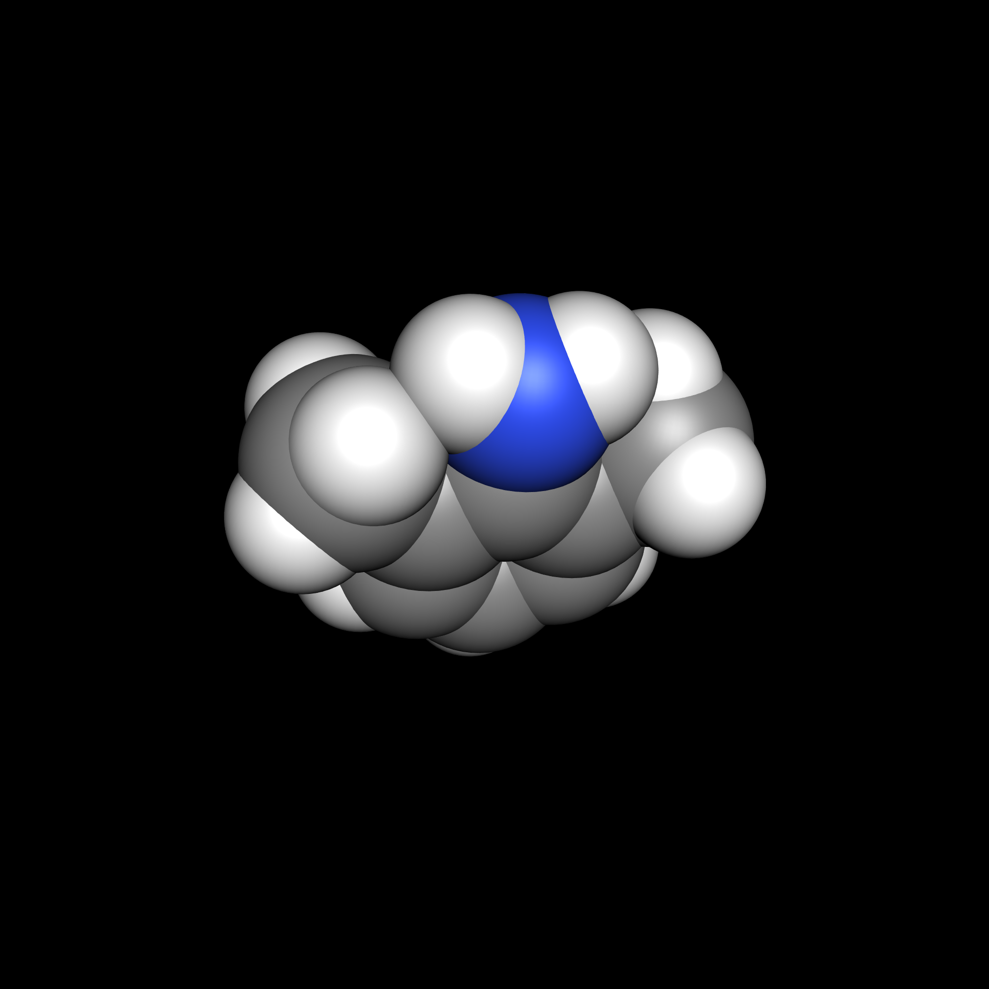
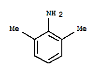
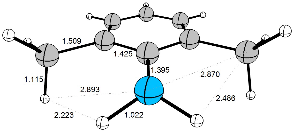
There are a number of different experimental parameters that you could check to attempt to find out more about 2,6-xylidine 1. I performed a few literature searches on different ones. For the remainder of this answer, xylidine is always taken to mean 2,6-xylidine.
Crystal structures
Naturally, the first idea of mine was to look up crytal structures. Unfortunately, xylidine is a liquid at room temperature, which may explain why I was unable to find a structure of the delinquent itself. Instead, I found a number of metal-coordinated xylidines. Hu, Mains and Holt investigated copper(I)-iodide clusters, and amoung them solved the structure of $\ce{[Cu4I4(MeCN)2(L)2]}$ 2 ($\ce{L} = \text{xylidine}$).[1] The structure is shown in figure 1.
![Structure of [Cu4I4(MeCN)2(L)2]](https://i.sstatic.net/J189j.png)
Figure 1: Structure of $\ce{[Cu4I4(MeCN)2(L)2]}$ 2 as solved by Hu, Mains and Holt.[1]
From their molecular analysis, $\ce{C20H28Cu4I4N4}$, it is evident that xylidine is a neutral (not anionic) ligand in this complex. The structure shows how the $\ce{N\bond{->}Cu}$ is almost perpendicular to the phenyl ring. This suggests a (possibly distorted since the positions of the hydrogen atoms are not determined) planar amino group as in the first of Martin’s structures.
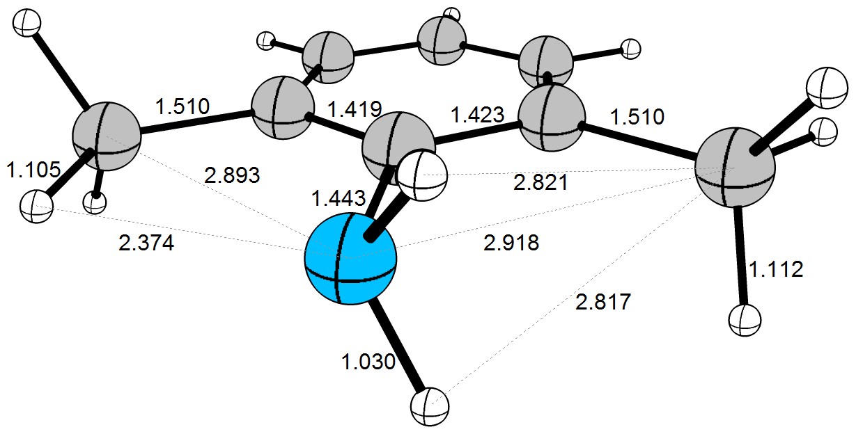
Humphries et al. studied niobium complexes and solved another few relevant structures.[2] In this answer, I will restrict myself to $\ce{[Nb({\eta^5-}C5H5)(CH3)(N-C6H3{-2,6-}Me2)(NH-C6H3{-2,6-}Me2)]}$ 3. This comlex features the parent xylidine structure both as an imido ligand ($\ce{R-N=M}$) and as an amido ligand ($\ce{R-HN-M}$). In this case, we are dealing with the anion of xylidine (one hydrogen abstracted). The structure is shown in figure 2.
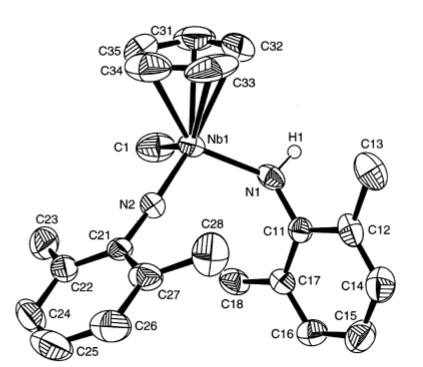
Figure 2: Structure of $\ce{[Nb({\eta^5-}C5H5)(CH3)(N-C6H3{-2,6-}Me2)(NH-C6H3{-2,6-}Me2)]}$ 3 as solved by Humphries et al.[2]
This structure, while a position of the xylidide hydrogen has been determined, has less informative value with respect to the configuration of the amino group: The oxidation state of niobium ($\mathrm{+V}$) is rather high making it rather small and the abstraction of a hydrogen means that an $\mathrm{sp^2}$-type hybrid orbital is used for coordination. Altogether, this means that this structure — while displaying an out-of-phenyl-plane lone pair — is less representative of xylidine and should be considered a representative of N-tert-butylxylidine instead. It should not be surprising that that molecule would adopt a perpendicular amino group rather than a planar one.
NMR shifts
After analysing crystal structures proved unsatisfactory, I turned my attention to NMR shifts. Both xylidine[3] 1 and aniline[4] 4 are featured in the spectral database of organic compounds (SDBS). The corresponding chemical shifts are given in table 1.
Table 1: NMR shifts of selected protons in xylidine 1 and aniline 4. Both compounds $0.04~\mathrm{ml}$ of amine in $0.5~\mathrm{ml}\ \ce{CDCl3}$. Spectra recorded at $90~\mathrm{MHz}$.[3,4]
\begin{array}{lccc}\hline
\text{compound} & \delta(\ce{NH2}) & \delta(\ce{{$m$-}CH}) & \delta(\ce{{$p$-}CH}) \\ \hline
\text{xylidine, }\textbf{1} & 3.46 & 6.93 & 6.62 \\
\text{aniline, }\textbf{4} & 3.55 & 7.12 & 6.73 \\ \hline \end{array}
In my opinion, these differences in chemical shift are minor. The slight upfield shift of the xylidine signals may, in my opinion, be attributed to the $+I$-effects of the two additional methyl groups which, according to theory, affect the meta protons most. Otherwise, hardly any difference can be observed. This seems to point to a structure of xylidine with a mostly planar amino group with respect to the phenyl ring.
$\mathrm{p}K_\mathrm{a}$ values
Finally, I decided to analyse the $\mathrm{p}K_\mathrm{a}$ values. The assumption here is that a lower $\mathrm{p}K_\mathrm{a}$ value indicates a greater stability of the unprotonated species which is most likely if the corresponding lone pair participates in resonance with the phenyl ring. The values are summarised in table 2.
$$\textbf{Table 2: }\mathrm{p}K_\mathrm{a}\text{ values of xylidine and selected related compounds.}\\
\begin{array}{lccc}\hline
\text{acid} & \text{base} & \mathrm{p}K_\mathrm{a} & \text{source}\\ \hline
\text{xylidine}\ce{. H+} & \textbf{1} & \phantom{0}3.90 & [5] \\
\text{aniline}\ce{. H+} & \textbf{4} & \phantom{0}4.87 & [6] \\
\ce{Me2HN^+-C6H5} & \textbf{5} & \phantom{0}5.07 & [7] \\
\ce{CH3NH3+} & \textbf{6} & 10.64 & [8] \\
\ce{(CH3)3NH+} & \textbf{7} & \phantom{0}9.81 & [9] \\
\ce{H2C=C(Me)-NHMe2+} & \textbf{8} & \phantom{0}8.28 & [10] \\ \hline \end{array}$$
Somewhat surprisingly, xylidine 1 has the lowest $\mathrm{p}K_\mathrm{a}$ value — surprising because a lower value should indicate a higher acidity which should indicate a better stabilised free base which does not fit with conjugation and the $+I$ effect of the methyl groups. However, we may also notice that the difference betweeeen xylidine 1 and aniline 4 is small, while methylamine 6 (which I chose in place of ammonia due to the presence of a carbon atom) differs by about $5{-}6$ logarithmic units making it substantially different. The significantly increased acidity of aniline 4 with respect to methylamine 6 is often explained with the delocalisation effect of the phenyl ring.
This opinion has, however, been contested. What we often fail to acknowledge when determining $\mathrm{p}K_\mathrm{a}$ values of phenyl-substituted heteroatoms is the significant difference in electronegativity between $\mathrm{sp^2}$ and $\mathrm{sp^3}$ carbons. Thus, one could say I am comparing apples and oranges when lumping methylammonium’s and anilinium’s $\mathrm{p}K_\mathrm{a}$ values into a single analysis. In fact, the difference between these two is lower than the difference between phenol and cyclohexanol with respect to the acidity constant.
For that reason, I included the seemingly unrelated N,N-dimethylaniline 5, trimethylamine 7 and 2-(dimethylamino)prop-1-ene 8 in the list of $\mathrm{p}K_\mathrm{a}$ values. Compound 8 features a nitrogen atom bound to one $\mathrm{sp^2}$-hybridised carbon and two $\mathrm{sp^3}$ hybridised ones. This is the same carbon types as 5 features with the exception that 5 also features a phenyl ring which is predicted to support delocalisation of the lone pair. Indeed, the difference in $\mathrm{p}K_\mathrm{a}$ values between the conjugate acids of 5 and 8 is significant — $3.2$ logarithmic units — which shows that resonance must feature an additional stabilising effect.
Conclusion
Crystal structures were badly unsatisfactory for determining the structure of xylidine 1. The NMR analysis suggests a strong structural homology to aniline 4. Most notably, however, the $\mathrm{p}K_\mathrm{a}$ values of the conjugate acids 1, 4, 5, 6 and 8 as summarised in table 2 show a clear trend which indicates that 1 should be structurally very similar to 4 featuring a delocalised p lone pair and not similar to 6 with a localised lone pair. The high acidity of 1’s conjugate base cannot be explained with an $\mathrm{sp^2}$ carbon alone. I thus am inclined to believe that xylidine’s amino group is complanar with respect to the phenyl ring and that the lone pair participates in the π system.
References:
[1]: G. Hu, G. J. Mains, E. M. Holt, Inorg. Chim. Acta 1995, 240, 559. DOI: 10.1016/0020-1693(95)04583-X.
[2]: M. J. Humphries, M. L. H. Green, R. E. Douthwaite, L. H. Rees, J. Chem. Soc., Dalton Trans. 2000, 4555. DOI: 10.1039/b007178l.
[3]: Spectral Database for Organic Compounds SDBS, 2,6-xylidine. SDBS-no. 1825. Spectrum no. 1825HSP-03-481.
[4]: Spectral Database for Organic Compounds SDBS, 2,6-xylidine. SDBS-no. 905. Spectrum no. 905HSP-03-391.
[5]: B. H. Asghar, Monatsh. Chemie 2008, 139, 1191. DOI: 10.1007/s00706-008-0913-5.
[6]: Wikipedia entry Aniline
[7]: I. Kaljurand, A. Kütt, L. Sooväli, T. Rodima, V. Mäemets, I. Leito, I. A. Koppel, J. Org. Chem. 2005, 70, 1019. DOI: 10.1021/jo048252w.
[8]: Wikipedia entry Methylamine.
[9]: Wikipedia entry Trimethylamine.
[10]: SciFinder entry for ‘1-Propen-2-amine, N,**N-dimethyl-’, CAS-number 22499-75-8. Acid-base constant predicted by Advanced Chemistry Development (ACD/Labs) Software V11.02 (© 1994–2016 ACD/Labs).

![Structure of [Cu4I4(MeCN)2(L)2]](https://i.sstatic.net/J189j.png)

